oct b scan
Die optische Kohärenztomographie ist ein bildgebendes Verfahren um 2- und 3-dimensionale Aufnahmen aus streuenden Materialien in Mikrometerauflösung zu erhalten. With the OCT3 system the number of A-scans per B-scan is 512.

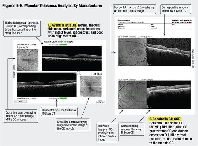
Oct B Scans Of The Retina Obtained With Different Imaging Techniques Download Scientific Diagram
Der andere Teil durchläuft eine Referenzstrecke.

. 1 Pieces Sourcing Request From. Sequential OCT B-scans The sequential examinations by OCT are acquired up to four times at the same location during a single examination. The advantage of this type of presentation B-Scan is that both the length of the flaw and its depth below the surface is revealed.
The horizontal axis on the scan connects to the probe position as it moves along the test object surface and gives information on the flaw. The edema can be seen as green extravascular signal extending across the foveal avascular zone. It is used for producing a two-dimensional cross-section of the eye and its orbit.
B-OCT was implemented in the spectral domain OCT device for posterior and anterior segment imaging REVO NX Optopol Technology. The location of this feature on OCT B-scans correlated to the sites of polypoidal lesions on ICGA. OCT-Angiography map These maps are a reconstruction of the microvasculature of the retina and.
CIRRUS 6000 is the next-generation OCTOCTA from ZEISS delivering high-speed image capture at 100000 scans per second with HD imaging detail and a wider field of view so clinicians can make more informed decisions and spend more time with their patients. OCT 10-mm B Scan LSO with B-scan location showing an averaged 10-mm B-scan image. However the U-shaped elevations were visible primarily before beginning treatment Dr.
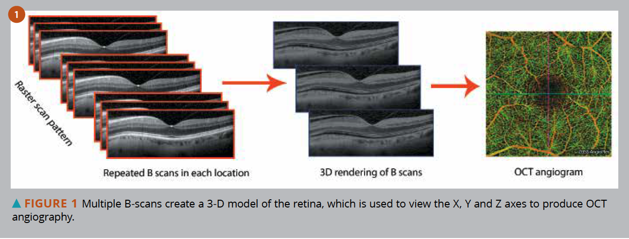
A total of 349 eyes 214 of healthy subjects 115 of patients with cataract and 20 with severe macular diseases were enrolled in the study. B-C B-scans extending through two areas of the hyper-reflective macular edema. Data processing OCT B-scan of the blood circulation Each group of OCT B-scan generates an image of the blood flow.
On the ultrasonic machine display a colored or gray scale intensity is used to display echo amplitude. Oct B-Scan Date Posted. Ein Teil wird auf die Probe gelenkt.
In the case of OCT2 each B-scan is based on 100 A-scans equally divided along the scan. 10 Product Description Buyer Business Info. A Color depth-encoded OCT angiogram of an eye with moderate non-proliferative diabetic retinopathy and exudative macular edema.
ZEISS CIRRUS 6000 ZEISS CIRRUS 5000. Dazu wird breitbandiges Licht von zeitlich geringer Kohärenzlänge in einem Strahlteiler in zwei Teile geteilt. A B-scan is generally used to evaluate diseases involving the posterior segment the hind two-third of the eye and orbit typically when the ocular media fluids within the eye are cloudy and a direct visualization is not possible.
We showed that if you only looked at the after-treatment OCT B-scan only a quarter of the eyes with PCV were diagnosed. The OCT scan uses a laser without radiation to obtain higher resolution images of the layers of the retina and optic nerve. OCT 6-mm B Scan LSO with B-scan locations showing a 4 other averaged 6-mm B.
ZD Medicals OCTA2020 system enables real-time scans of the macula optic disc and retina. The results of B-OCT were compared to swept source OCT-based IOLMaster 700 Carl. An OCT eye exam is a non-invasive test that provides 3-D color-coded cross sectional images of the retina to enable early detection and treatment of ocular disease that may develop without any noticeable symptoms.
Automated Segmentation of. C Ground truth map with three categories background green retinal tissue black and retinal fluid area red. But if you looked at the before-treatment B.
Looking to purchase an OCT as well as a B-Scan unit. Thus for a given width of the scan line the OCT3 measurement provides a smaller transverse pixel spacing than is obtained with the OCT2. Overlaying flow signal red over the.
B-scan is considered the brightness scan.

Explaining Oct Scans With Their Mechanism And Benefits

Oct Scanning Modes A A Line B B Scan C Volumetric Oct The Oct Download Scientific Diagram
Classification Of Healthy And Diseased Retina Using Sd Oct Imaging And Random Forest Algorithm Plos One

Layer By Layer B Scan Sd Oct Display Of Normal Retina Abbreviation Download Scientific Diagram

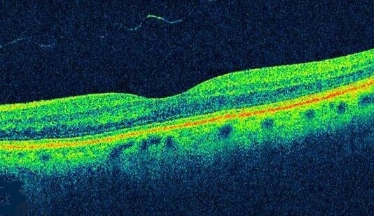
Figure 12 2 Oct B Scan Of The Retina Brighter Pixels Indicate Tissue Which Reflects More Light The Upper Portion Of The Figure Shows The Vitreous Humor Which Has Very Low Reflectivity The Small

Fig 3 19 Ps Oct B Scan Through The Fovea High Resolution Imaging In Microscopy And Ophthalmology Ncbi Bookshelf
Diabetic Retinopathy Optical Coherence Tomography Scans

3 Oct B Scan Image Robert Lloyd

Oct Scanning And Scanner Coordinate System Schematic Left 1d Download Scientific Diagram

What Is Oct And How Can It Help Ophthalmologists Acquire High Resolution Information On Ocular Tissue Science Lab Leica Microsystems

Layer By Layer B Scan Sd Oct Display Of Normal Retina Abbreviation Download Scientific Diagram

Intra Retinal Layer Segmentation Of Oct B Scans A Exemplary B Scan Download Scientific Diagram
Exudates Optical Coherence Tomography Scans

Comparison Of Pseudo Slo Images Oct B Scan Images And The Download Scientific Diagram


Comments
Post a Comment